
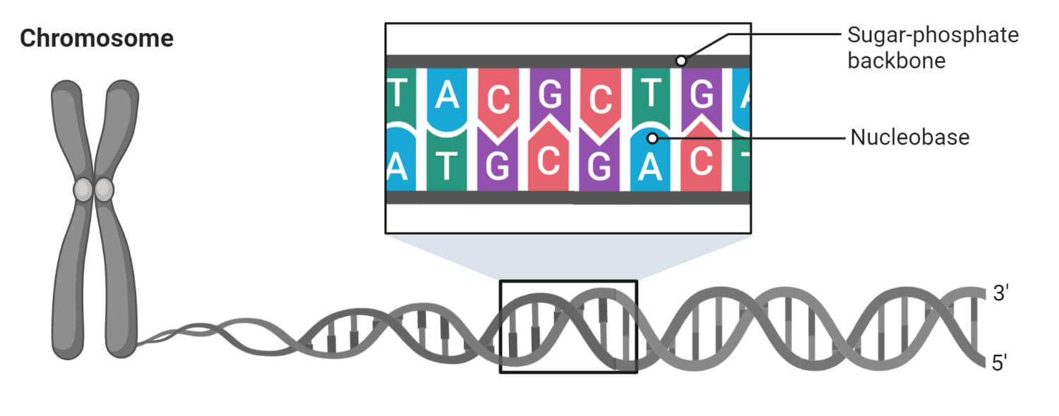
What are Chromosomes?
In thenucleusof each cell, theDNAmolecule is packaged into thread-like structures called chromosomes. Each chromosome is made up of DNA tightly coiled many times around proteins called histones that support its structure. Chromosomes were first described by Strasburger (1815), and the term ‘chromosome’ was first used by Waldeyer in 1888. They appear as rod-shaped dark stained bodies during the metaphase stage ofmitosiswhen cells are stained with a suitable basic dye and viewed under alight microscope.
- Chromosomesare the nuclear components of the special organization, individuality, and function that are capable of self-reproduction and play a vital role in heredity, mutation, variation and evolutionary development of the species.
- Each chromosome is made up ofDNAtightly coiled many times around proteins that support its structure.
- The proteins that bind to the DNA to form eukaryotic chromosomes are traditionally divided into two classes: the histones and the non-histone chromosomal proteins.
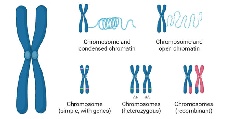
- The complex of both classes of protein with the nuclear DNA of eukaryotic cells is known as chromatin.
- Chromatin are a highly compacted structure consisting of packaged DNA and necessary so as to fit DNA into the nucleus.
- The assembly of DNA into chromatin involves a range of events, beginning with the formation of the basic unit, the nucleosome, and ultimately giving rise to a complex organization of specific domains within the nucleus.
- In the first step of this process, DNA is condensed into an 11 nm fiber that represents an approximate 6-fold level of compaction. This is achieved through nucleosome assembly.
- The nucleosome is the smallest structural component of chromatin and is produced through interactions between DNA and histone proteins.
- Each nucleosome consists of histone octamer core, assembled from the histones H2A, H2B, H3 and H4 (or other histone variants in some cases) and a segment of DNA that wraps around the histone core. Adjacent nucleosomes are connected via “linker DNA”.
Structure of a Chromosome
- In eukaryotes the chromosomes are multiple large, linear and are present in the nucleus of the cell.
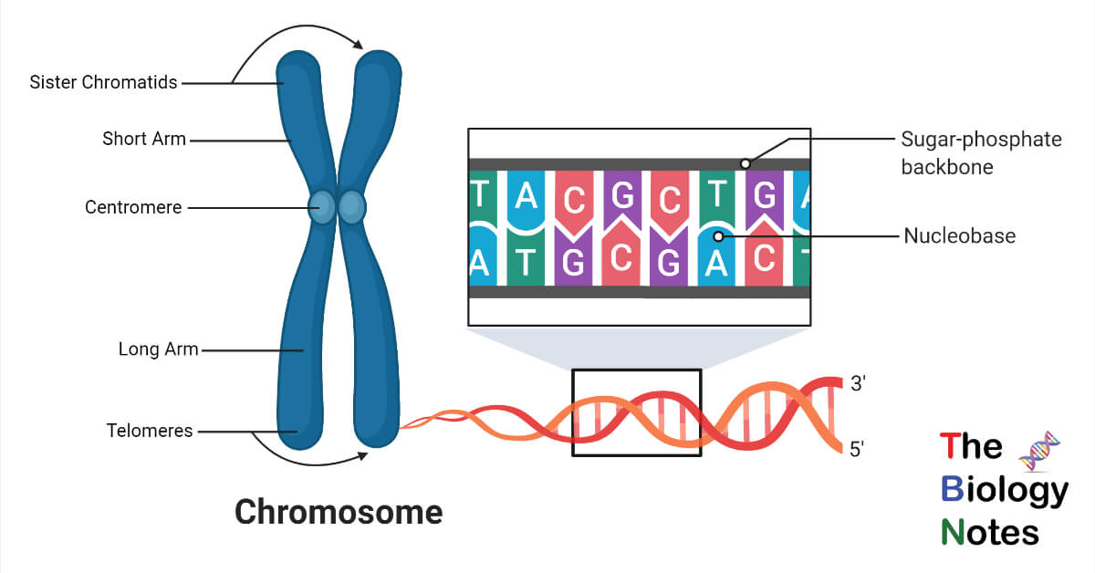
- Each chromosome typically has one centromere and one or two arms that project from the centromere.
- Structurally, each chromosome is differentiated into three parts—
- Pellicle
- Matrix
- Chromonemata
Pellicle
- It is the outer envelope around the substance of chromosome.
- It is very thin and is formed of achromatic substances.
Matrix
- It is the ground substance of chromosome which contains the chromonemata.
- It is also formed of non-genic materials.
Chromonemata
- Embedded in the matrix of each chromosome are two identical, spirally coiled threads, the chromonemata.
- The two chromonemata are also tightly coiled together that they appear as single thread of about 800A thickness.
- Each chromonemata consists of about 8 microfibrils, each of which is formed of a double helix of DNA.
In mitotic metaphase chromosomes, the following structural feature (except chromomere) can be seen under the light microscope:
(1) Chromatid,
(2) Chromonema,
(3) Chromomeres,
(4) Centromere,
(5) Secondary constriction or Nucleolar organizer,
(6) Telomere and
(7) Satellite.
Centromere
- A small structure in the chromonema, marked by a constriction which is recognised as permanent structure in the chromosome is termed as the centromere.
- At this point the two chromonemata are joined together.
- It is known as centromere or kinetochore or primary constriction.
- 它将染色体划分为两个部分,或“rms.” The short arm of the chromosome is labeled the “p arm.” The long arm of the chromosome is labeled the “q arm.”
- Its position is constant for a given type of chromosome and forms a feature of identification.
- In thin electron microscopic sections, the kinetochore shows a trilaminar structure, i.e., a 10 nm thick dense outer protein aceous layer, a middle layer of low density and a dense inner layer tightly bound to the centromere.
- The chromosomes are attached to spindle fibres at this region during cell division.
Secondary Constriction or Nucleolar Organiser
- 染色体除了主缩tion or the centromere possesses secondary constriction at any point of the chromosome.
- Constant in their position and extent, these constrictions are useful in identifying particular chromosomes in a set.
- The chromosome region distal to the secondary constriction i.e., the region between the secondary constriction and the nearest telomere is known as satellite.
- Therefore, chromosomes having secondary constrictions are called satellite chromosomes or sat-chromosomes.
- Nucleolus is always associated with the secondary constriction of sat-chromosomes. Therefore, secondary constrictions are also called nucleolus organiser region (NOR) and sat-chromosomes are often referred to as nucleolus organiser chromosomes.
Telomeres
- These are specialized ends of a chromosome which exhibits physiological differentiation and polarity.
- Each extremity of the chromosome due to its polarity prevents other chromosomal segments to be fused with it. The chromosomal ends are known as the telomeres.
- If a chromosome breaks, the broken ends can fuse with each other due to lack of telomere.
Types of Chromosomes
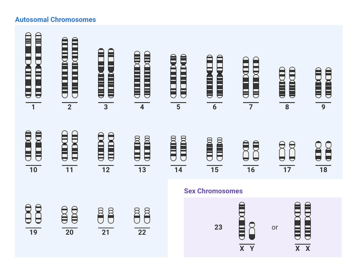
A. Autosomes and Sex Chromosomes
- Human chromosomes are of two types- autosomes and sex chromosomes.
- Genetic traits that are linked to the sex of the person are passed on through the sex chromosomes. The rest of the genetic information is present in the autosomes.
- 人类有23对染色体的细胞, of which 22 pairs are autosomes and one pair of sex chromosomes, making a total of 46 chromosomes in each cell.
B. On the Basis of Number of Centromeres
- Monocentricwith one centromere.
- Dicentricwith two centromeres.
- Polycentricwith more than two centromeres
- Acentric着丝粒。这种染色体代表联邦shly broken segments of chromosomes which do not survive for long.
- Diffused or non-locatedwith indistinct centromere diffused throughout the length of chromosome.
C. On the Basis of Location of Centromere
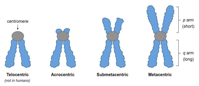
- Telocentricare rod-shaped chromosomes with centromere occupying the terminal position, so that the chromosome has just one arm.
- Acrocentricare also rod-shaped chromosomes with centromere occupying a sub-terminal position. One arm is very long and the other is very short.
- Sub-metacentricchromosomes are with centromere slightly away from the mid-point so that the two arms are unequal.
- Metacentricare V-shaped chromosomes in which centromere lies in the middle of chromosome so that the two arms are almost equal.
Prokaryotic Chromosomes
- The DNA of a bacterial cell, such asEscherichia coli, is a circular double-stranded molecule often referred to as the bacterial chromosome.
- The circular DNA is packaged into a region of the cell called the nucleoid where it is organized into 50 or so loops or domains that are bound to a central protein scaffold, attached to the cell membrane.
- The DNA is negatively supercoiled, that is, it is twisted upon itself.
- It is complexed with several DNA-binding proteins, the most common of which are proteins HU, HLP-1 and H-NS. These are histone-like proteins.
Eukaryotic Chromosomes
- The large amount of genomic DNA in a eukaryotic cell is tightly packaged in chromosomes contained within a specialized organelle, the nucleus.
- With the exception of the sex chromosomes, diploid eukaryotic organisms such as humans have two copies of each chromosome, one inherited from the father and one from the mother.
- Chromosomes contain both DNA and protein.
- Most of the protein on a weight basis is histones, but there are also many thousands of other proteins found in far less abundance and these are collectively callednon- histone proteins (NHP).
- This nuclear DNA–protein complex is called chromatin.
- In the nucleus, each chromosome contains a single linear double-stranded DNA molecule.
- The length of the packaged DNA molecule varies. In humans, the shortest DNA molecule in a chromosome is about 1.6 cm and the longest is about 8.4 cm.
- The extensive packaging of DNA in chromosomes results from three levels of folding involving nucleosomes, 30 nm filaments and radial loops.
a. Nucleosomes
- The first level of packaging involves the binding of the chromosomal DNA to histones.
- Overall, in chromosomes, the ratio of DNA to histones on a weight basis is approximately 1:1.
- There are five main types of histones called H1, H2A, H2B, H3 and H4.
- Histones are very basic proteins; about 25% of their amino acids are lysine or arginine so histones have a large number of positively charged amino acid side-chains.
- These positively charged groups therefore bind to the negatively charged phosphate groups of DNA
b. 30 nm fiber
- If nuclei are lysed very gently, the chromatin is seen to exist as a 30 nm diameter fiber.
- 纤维是由组蛋白H1分子痣ng to the linker DNA of each nucleosome at the point where it enters and leaves the nucleosome.
- The histone H1 molecules interact with each other, pulling the nucleosomes together.
c. Radial loops
- When chromosomes are depleted of histones, they are seen to have a central fibrous ‘protein scaffold’ (or nuclear matrix) to which the DNA is attached in loops.
- Therefore, in vivo it seems likely that the next order of packaging involves the attachment of the 30 nm fiber to multiple locations on this central protein scaffold in a series of radial loops.
- The mitochondria and chloroplasts of eukaryotic cells also contain DNA but, unlike the nuclear DNA, this consists of double-stranded circular molecules resembling bacterial chromosomes.
Nucleosome Model of Chromosome
- Nucleosome model is a scientific model which explains the organization of DNA and associated proteins in the chromosome.
- It also further explains the exact mechanism of the folding of the DNA in the nucleus.
- The model was proposed by Roger Kornberg in 1974 and is the most accepted model of chromatin organization.
- It was confirmed and christened by P. Oudet et al., (1975).
Features of the Nucleosome Model of Chromosomes
- In eukaryotes, DNA is tightly bound to an equal mass of histones, which serve to form a repeating array of DNA-protein particles, called nucleosomes.
- If it was stretched out, the DNA double-helix in each human chromosome would span the cell nucleus thousands of time.
- Histones play a crucial role in packing this very long DNA molecule in an orderly way (i.e., nucleosome) into nucleus only a few micrometers in diameter.
- Thus, nucleosomes are the fundamental packing unit particles of the chromatin and give chromatin a “beads-on-a-string” appearance in electron micrographs taken after treatments that unfold higher-order packing.
- Each nucleosome is a disc-shaped particle with a diameter of about 11 nm and 5.7 nm in height containing 2 copies of each 4 nucleosome histones–H2A, H2B, H3, and H4.
- This histone octamer forms a protein core [(i.e., a core of histone tetramer (H3, H4)2 and the apolar regions of 2(H2A and H2B)] around which the double-stranded DNA helix is wound 1¾ time containing 146 base pairs.
- In chromatin, the DNA extends as a continuous thread from nucleosome to nucleosome.
- Each nucleosome bead is separated from the next by a region of linker DNA which is generally 54 base pair long and contains single H1 histone protein molecule.
- Generally, DNA makes two complete turns around the histone octamers and these two turns (200 bp long) are sealed off by H1 molecules.
- On average, nucleosomes repeat at intervals of about 200 nucleotides or base pairs. For example, a eukaryotic gene of 10,000 nucleotide pairs will be associated with 50 nucleosomes and each human cell with 6 x 10⁹DNA nucleotide pairs contains 3 x 10⁷
The Folding of the DNA
- The first step is the assembly of the DNA with a newly synthesized tetramer (H3-H4), are specifically modified (e.g. H4 is acetylated at Lys5 and Lysl2 (H3-H4)), to form a sub-nucleosomal particle, which is followed by the addition of two H2A-H2B dimers.
- This produces a nucleosomal core particle consisting of 146 base pairs of DNA bind around the histone octamer. This core particle and the linker DNA together form the nucleosome.
- The next step is the maturation step that requires ATP to establish regular spacing of the nucleosome cores to form the nucleo-filament.
- During this step the newly incorporated histones are de-acetylated.
- Next, the incorporation of linker histones is accompanied by folding of the nucleo-filament into the 30 nm fiber, the structure of which remains to be elucidated.
- Two principal models exist- the solenoid model and the zig-zag.
- Finally, further successive folding events lead to a high level of organization and specific domains in the nucleus.
Function and Significance of Chromosomes
- The number of the chromosomes is constant for a particular species. Therefore, these are of great importance in the determination of the phylogeny and taxonomy of the species.
- Genetic Code Storage: Chromosome contains the genetic material that is required by the organism to develop and grow. DNA molecules are made of chain of units called genes. Genes are those sections of the DNA which code for specific proteins required by the cell for its proper functioning.
- Sex Determination:Humans have 23 pairs of chromosomes out of which one pair is the sex chromosome. Females have two X chromosomes and males have one X and one Y chromosome. The sex of the child is determined by the chromosome passed down by the male. If X chromosome is passed out of XY chromosome, the child will be a female and if a Y chromosome is passed, a male child develops.
- Control of Cell Division:Chromosomes check successful division of cells during the process of mitosis. The chromosomes of the parent cells insure that the correct information is passed on to the daughter cells required by the cell to grow and develop correctly.
- Formation of Proteins and Storage:The chromosomes direct the sequences of proteins formed in our body and also maintain the order of DNA. The proteins are also stored in the coiled structure of the chromosomes. These proteins bound to the DNA help in proper packaging of the DNA.
The Human Chromosomes
- Every normal human cell, except for sperm and egg cells, has 23 pairs of chromosomes for a total of 46 chromosomes.
- Sperm and egg cells have only one of each pair of chromosomes for a total of 23. Each chromosome contains hundreds to thousands of genes.
- The sex chromosomes are one of the 23 pairs of chromosomes. Normal people have 2 sex chromosomes, and each is either an X or a Y chromosome. Normal females have two X chromosomes (XX), and normal males have one X and one Y chromosome (XY).
Chromosome Abnormalities
Chromosomal abnormalities can fall under two categories:
- Structural abnormalities(eg, deletions or rearrangements) or
- Abnormalities of chromosomal number.
Abnormalities of chromosomal number generally arise from meiotic non- disjunction (failure of chromosome pairs to separate during cell division) or through anaphase lag (loss of chromosome during cell division).
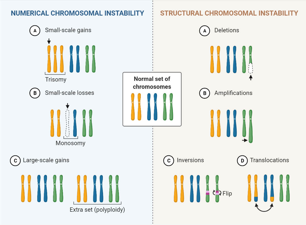
Some common chromosomal disruptions include:
- Aneuploidy:Chromosome number that is not a multiple of 23.
- Polyploidy:Chromosome number that is 3 or 4 times the haploid number of 23.
- Deletion:Loss of part of chromosome.
- Translocation:Exchange of chromosome parts between non-homologous chromosomes.
- Balanced translocation:No genetic material lost; clinically asymptomatic.
- Robertsonian translocation:Joining of long arms of two acrocentric chromosomes with loss of short arms.
- Inversion:Reunion of separated portion.
Some Important Chromosomal Disorders
A. Cri du Chat Syndrome
Cause
Caused by partial deletion of chromosome 5p.
Characteristics
Patients exhibit a high-pitched cat-like cry at birth, which is usually due to structural abnormalities in the larynx; patients also have severe developmental delay and cognitive deficits and distinctive facial abnormalities (round face, low-set ears, microcephaly, and a hypoplastic nasal bridge).
Lab findings: Cytogenetic studies reveal a deletion of chromosome 5p.
B. Down Syndrome
Cause
Trisomy 21 causes 95% of cases (usually because of meiotic nondisjunction); 4% of cases are caused by Robertsonian translocation of the long arm of chromosome 21 to another chromosome (usually chromosome 14 or 22); 1% of cases are caused by mosaicism, resulting from mitotic nondisjunction of chromosome 21 during embryogenesis.
Characteristics
Severe mental retardation;duodenal and esophageal atresia; short hands with simian crease (single palmar crease). Specific facial features include flat face; epicanthal folds; wide-set eyes; Brushfield spots (white spots on periphery of iris).
Congenital heart defects: Endocardial cushion defects leading to ostium primum atrial septal defects, ventricular septal defects, and atrioventricular valve malformations.
C. Edward Syndrome
Cause
Most cases are caused by trisomy 18 (usually caused by meiotic nondisjunction). A few cases are caused by mosaicism, resulting from mitotic nondisjunction of chromosome 18 during embryogenesis.
Characteristics
Severe mental retardation; rocker-bottom feet; specific facial features include prominent occiput, micrognathia (small jaw), low-set ears; congenital heart and renal defects; overlapping third and fourth fingers.
D. Klinefelter syndrome
Cause
This disorder is characterized by two or more X chromosomes with one or more Y chromosomes (most commonly 47, XXY karyotype with a single Barr body) and is most commonly caused by maternal meiotic nondisjunction. Other causes include mosaicism or paternal meiotic nondisjunction.
Characteristics
Small atrophic testes; tall stature; lack of secondary male characteristics and gynecomastia.
Male infertility, often resulting from reduced spermatogenesis. Occasionally associated with mild mental retardation.
Lab findings: Decreased testosterone levels; increased FSH and LH levels.
E. Patau Syndrome
Cause
Most cases are caused by trisomy 13 (usually caused by meiotic nondisjunction). A few cases are caused by mosaicism, resulting from mitotic nondisjunction of chromosome 13 during embryogenesis or translocation between chromosomes 13 and 14.
Characteristics
Severe mental retardation; microcephaly and holoprosencephaly; cleft lip and palate and microphthalmia (small eyes); polydactyly; congenital heart and renal defects; umbilical hernia; rocker-bottom feet.
F. Prader-Willi syndrome and Angelman syndrome
Cause
Both diseases are due to an identical deletion on chromosome 15q and demonstrate imprinting, a phenomenon in which the same mutation results in different phenotypes depending on whether the mutated chromosome was of maternal or paternal origin.
Prader-Willi syndrome develops when the deletion is on the paternally derived chromosome, whereas Angelman syndrome develops when the deletion is on the maternally derived chromosome.
Characteristics
Prader-Willi syndrome: Mental retardation; hypogonadism; hypotonia; obesity leading to diabetes.
Angelman syndrome: “Happy puppet” with ataxic gait and inappropriate laughter; mental retardation; seizures.
G. Turner syndrome
Caused by partial or complete monosomy of the X chromosome (XO karyotype with no (Barr body).
Characteristics
Short stature with a broad chest and widely spread nipples; cystic hygroma of the neck, leading to webbed-neck appearance; lymphedema of extremities; coarctation of the aorta and other congenital heart defects.
Reproductive symptoms include primary amenorrhea, replacement of ovaries with fibrous strands (no ova or follicles), and infantile genitalia and breasts.
Lab findings: Decreased estrogen production; increased FSH and LH levels.
References
- Wigler MH, Axel R. Nucleosomes in metaphase chromosomes.Nucleic Acids Res. 1976;3(6):1463-1471. doi:10.1093/nar/3.6.1463
- David Hames and Nigel Hooper (2005). Biochemistry. Third ed. Taylor & Francis Group: New York.
- Verma, P. S., & Agrawal, V. K. (2006). Cell Biology, Genetics, Molecular Biology, Evolution & Ecology (1 ed.). S .Chand and company Ltd.
- McGinty, R. K., & Tan, S. (2014). Nucleosome structure and function.Chemical reviews,115(6), 2255– doi:10.1021/cr500373h
- Alberts, B., Johnson, A., Lewis, J., Raff, M., Roberts, K., & Walter, P. (2002). Molecular biology of the cell. New York: Garland Science.
- Baron J.S. and Lee I.C. Biochemistry & Genetics. Second Edition McGraw Hill Medical: New York.
- http://www.yourarticlelibrary.com/biology/chromosomes-structure-functions-and-other-details-about-chromosomes/22946
- https://medlineplus.gov/genetics/understanding/basics/chromosome/
- https://www.britannica.com/science/chromosome
- https://biology.tutorvista.com/cell/chromosomes.html
- https://www.easybiologyclass.com/nucleosome-model-of-chromosomes-in-eukaryotes-short-notes/
- http://www.biologydiscussion.com/cell-biology/nucleosome-model/nucleosome-model-of-chromatin-assembly-cell-nucleus-biology/78886
- https://www.mechanobio.info/genome-regulation/what-are-nucleosomes/
- https://www.msdmanuals.com/home/fundamentals/genetics/genes-and-chromosomes
- https://www2.le.ac.uk/projects/vgec/highereducation/topics/dna-genes-chromosomes.
- https://www.merckmanuals.com/home/children-s-health-issues/chromosome-and-gene-abnormalities/overview-of-chromosome-and-gene-disorders
- https://www.merriam-webster.com/dictionary/chromosome