
Amicroscopeis an optical instrument having one or more lenses system which is used to get a clear magnified image of minute objects or structures that can’t be viewed by the naked eyes.
Derived from Greek words“mikrós” meaning “small” and“skópéō”meaning “look at”.
They are devices used to observe the detailed structure of small objects. Very small objects that can’t be seen by unaided eyes like cells, microorganisms, viruses, nanoparticles, sub-cellular structures, etc. are viewed by using a microscope. Such objects that can be viewed by using a microscope only are called ‘microscopic’.
There are different types of microscopes like a light microscope, dark-field microscope, phase contrast microscope, electron microscope, fluorescent microscope, etc.
Broadly parts of a microscope can be studied in 2 groups; optical parts, including lenses and light source, and structural parts, including head, base, arms, and joints. Modern microscopes have additional electronics and display devices.
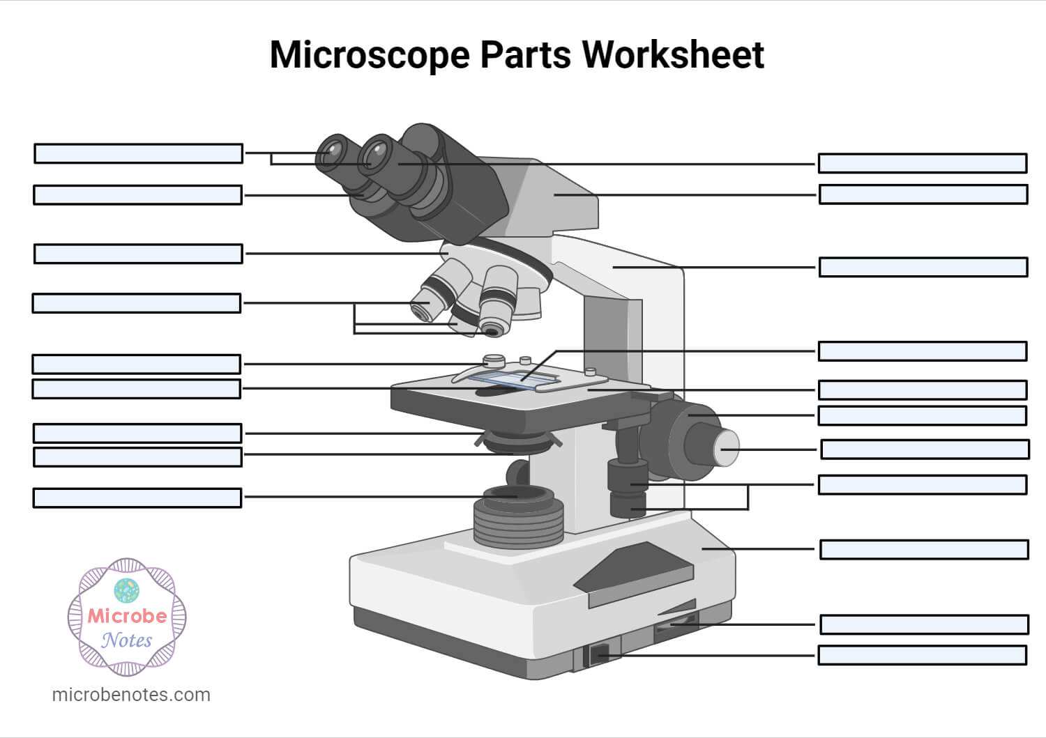
Microscope Parts Worksheet
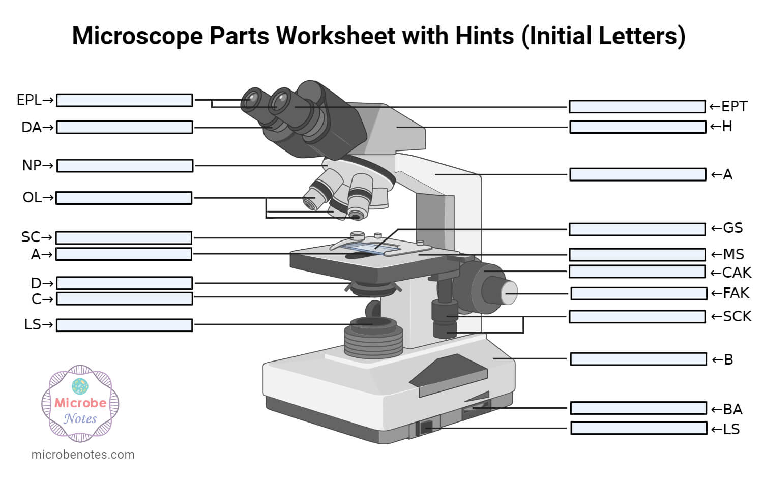
Microscope Parts Worksheet Hints
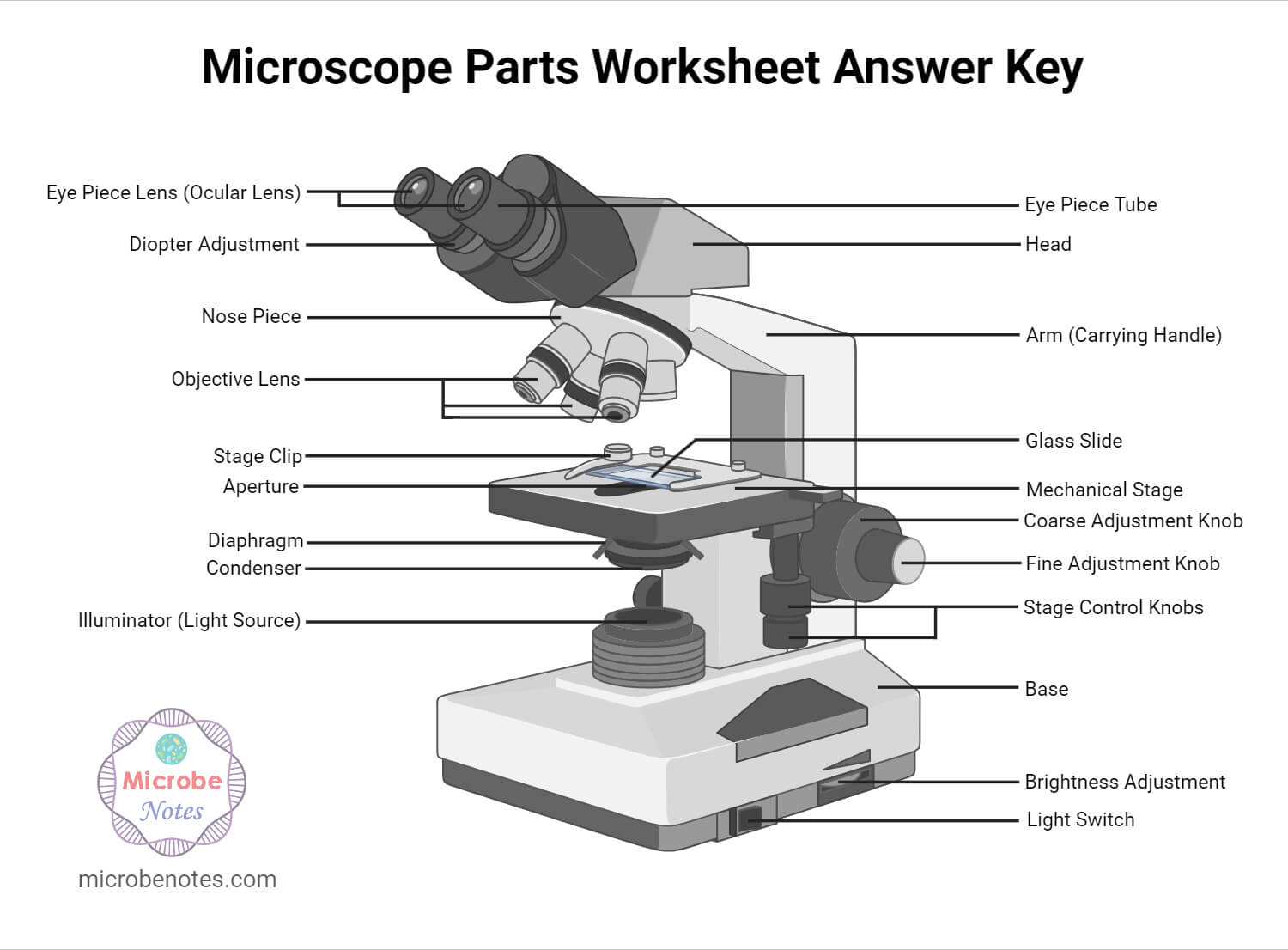
Microscope Parts Worksheet Answer Key
Download Microscope Parts Worksheet PDF
List of 17 parts of a microscope
- Illuminator (Light Source)
- Diaphragm (Iris)
- Condenser
- Condenser Focus Knob
- Rack Stop
- Stage
- Stage Control Knobs
- Nose Piece
- Objective Lens
- Tube (Head)
- Eyepiece (Ocular Lens)
- Diopter Adjustment
- Adjustment Knobs
a. Fine Adjustment Knob
b. Coarse Adjustment Knob - Arm
- Base
- Light Switch
- Brightness Adjustment
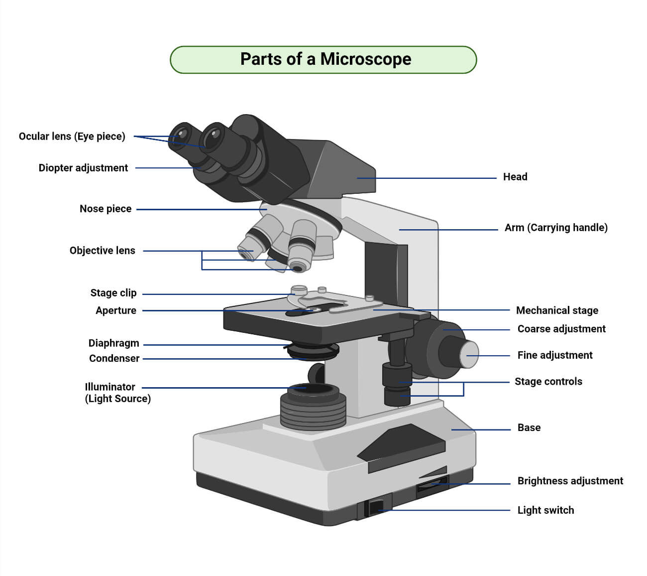
A typical microscope contains the following parts;
1. Illuminator (Light Source)
Amicroscopic illuminatoris a light source. In some compound microscope, the mirror is used which reflect the light from an external source to the sample. In other optical microscopes, different electric bulbs of low voltages are used as a constant light source. Commonly used illuminators are tungsten-halogen lamps, 75-150W Xenon lamps, tin-halide lamps, mercury vapor lamps, etc. The selection of types of a bulb is based on the requirement of intensity and wavelength for illumination.
2. Diaphragm (Iris)
Diaphragm (Iris)is an adjustable apparatus that regulates the amount of light that reaches the specimen. It is located below the stage, just above the illuminator. It regulates the intensity and size of the beam of light striking the specimen.
3. Condenser
A condenseris a lens system that collects and focuses the light rays coming from the illuminator on the specimen being viewed. It is found below the stage just above the condenser. It is useful while working at a high magnification of 400X or above. It renders a clearer and sharper image than those without a condenser lens in high magnification.
In modern and high-quality microscopes, an Abbe condenser is used. It is moveable and can move towards or away from the stage. It controls the diameter of the beam of light striking the specimen and regulates the brightness, illumination, and contrast of the developing image.
4. Condenser Focus Knob
Condenser Focus Knobis an adjustment knob that moves the condenser up and down. It helps in controlling the focus of the light passing from the condenser to the specimen.
5. Rack Stop
Rack Stopis another adjustment device that regulates how close the stage can rise without hitting the objective lens. It prevents the specimen slide from hitting the objective lens and damaging the specimen as well as the lens.
6. Stage
The stageis a flat platform where a slide with a specimen is placed for viewing. The stage has a central aperture through which the focused light from the condenser strikes the specimen. It contains two-stage clips for holding the slide in a fixed position.
Most microscopes have a mechanical stage. This type of stage allows for the mechanical movement of slides using stage control knobs.
7. Stage Control Knobs
Stage Control Knobsare the control knobs used to move the stage mechanically. There are two knobs; one for moving left and right, and the other for moving forward and backward. This will move the slide in the field of vision.
8. Nose Piece
Nose Piece是一个可移动的圆形结构,房屋the objective lenses. It is also called the revolving turret. It is connected to the body tube and lies just above the stage. It can be rotated clockwise or counterclockwise to increase or decrease the magnification respectively. The change in magnification results due to a change in the objective lens.
9. Objective Lens
Objective Lensis the lenses that are closest to the specimen. They are fitted on the nose piece. In a standard microscope, there are 3 to 4 objective lenses of different magnifying power viz. 4X, 10X, 40X and 100X. The objective lenses first receive the light transmitted from the specimen and magnify the image for the first time.
Objective lenses are color-coded and are of different sizes. Size and color depend on the power of the lens. The smallest lens is of the lowest power and gradually the longest will be of the highest power. The high power lenses i.e. 40X and 100X are retractable i.e. their end can be pushed inward. In most optical microscopes, objective lenses with 100X or more magnification are of oil immersion type.
10. Tube (Head)
Tube (Head)is a cylindrical metallic tube that holds the eyepiece lens at one end and connects to the nose piece at other ends. It is also called a body tube or eyepiece tube. It connects the eyepiece lens to the objective lens. The light coming from objectives will bend inside this tube.
In binocular microscopes, they are adjustable so that the viewer can adjust the eyepiece for maximum visualization.
11. Eyepiece (Ocular Lens)
Eyepiece (Ocular Lens)are the lenses that are closest to the viewer’s eye. They are located at the top of the microscope. This part is used to look at the specimen.
These lenses come in different magnification power from 5X to 30X, but the most common ocular lenses are of 10X or 15X magnification.
They magnify the image for the second time.
12. Diopter Adjustment
Diopter Adjustmentis a control knob present only in the binocular microscope that is used to change focus on one eyepiece. It is used to correct any difference in vision and compensate for the differences in vision between the two eyes of the viewer.
13. Adjustment Knobs
Adjustment Knobsare the control knobs used to focus the microscope on the specimen. These knobs are of two types;
a. Fine Adjustment Knob
Fine Adjustment Knobis used for fine adjustment. It is a smaller knob and is used to move the stage up or down very slowly. A very small distance is covered by the stage on each rotation of the fine adjustment knob. It is used to sharpen the image. It is mostly used while viewing under high power.
b. Coarse Adjustment Knob
Coarse Adjustment Knobis used for focusing the image under low power magnification. It is a larger knob and is used to move the stage up or down very rapidly. The stage is raised or lowered rapidly with the help of a coarse adjustment knob.
14. Arm
Armis a structural part of the microscope that connects the head (tube) with the base of the microscope. It provides support to the head.
15. Base
Baseis the lowermost part of the microscope that supports the entire microscope structure. It provides stability for the microscope. Illuminator, light switches, and electrical wiring system are fitted in the base.
16. Light Switch
Light Switchis an electrical control device. Light switches are used to on and off the illuminator.
17. Brightness Adjustment
The brightness adjustment system controls the voltage supplied to the light bulb controlling the intensity (brightness) of the light bulb.
How Does Microscope Work?
In a simple light microscope, a thin specimen containing a slide is placed on the microscope’s stage.
A beam of light is passed through the condenser to the specimen. The light transmitted from the specimen enters the objective lens. While passing through the objectives, the transmitted rays are spread so that they appear to come from the bigger objects.
The light is then focused on the eyepiece lens. This lens further magnifies the pre-magnified image coming from the objectives.
Finally, a highly magnified image can be observed.
References
- microscope | Types, Parts, History, Diagram, & Facts | BritannicaParts of the Microscope with Labeling (also Free Printouts) – Laboratoryinfo.com
- 的部分(microbio显微镜及其功能logynote.com)
- Parts of a Microscope – The Comprehensive Guide – Microscope and Laboratory Equipment Reviews (microscopespot.com)
- Microscope: Definition, Types, Uses, Parts & Examples | Toppr
- Simple Microscope Definition, Magnification, Parts And Uses (byjus.com)
- 相衬显微镜——定义,原则,Parts, Uses (learninsta.com)
- What is FluorescenceMicroscopy? Definition, Principle, Fluorescence & Parts – Biology Reader
- Electron Microscope: Definition, Types, Parts, Application, Advantages, Disadvantages (microbiologynote.com)
- DARK FIELD MICROSCOPE: CHARACTERISTICS, PARTS, FUNCTIONS – SCIENCE (warbletoncouncil.org)
- Dissecting Stereo Microscope Parts and Functions (microscopemaster.com)
- Confocal Microscope Principle, Uses, Parts, Advantages, and Disadvantages. (microbiologynote.com)
- Inverted microscope Images and definition, principle, Uses, Parts. (microbiologynote.com)
- USB Microscope- definition, principle, parts, examples, uses.docx – USB Microscope- definition, principle, parts, examples, uses March 26, 2020 by Faith | Course Hero